 Write to the booksellers
-
Type
Book (2300)
Magazine (8)
-
Latest
Last 24h (1)
Last 3 days (3)
Last month (8)
Last week (2)
-
Language
English (1)
French (2305)
Latin (2)
-
Century
16th (4)
17th (17)
18th (97)
19th (326)
20th (782)
21st (99)
-
Countries
Belgium (115)
Canada (1)
Côte d'Ivoire (2)
Denmark (1)
France (1783)
Greece (1)
Italy (36)
Switzerland (369)
-
Syndicate
ALAC (1)
CLAM (1)
ILAB (1291)
NVVA (163)
SLACES (163)
SLAM (944)
SNCAO (3)
PRECIS D'ANATOMIE - ANATOMIE DES MEBRES OSTEOLOGIE DU CRANE, DE LA FACE, DU THORAX, DU BASSIN - BIBLIOTHEQUE DU DOCTORAT EN MEDECINE - TOME 1
J.B. BAILLIERE. 1948. In-8. Broché. Etat passable, Livré sans Couverture, Dos abîmé, Papier jauni. 318 Pages. Nombreuses figures en couleur et en noir et blanc dans et hors texte. Dos abimé. Quelques rousseurs sans conséquence pour la lecture. Absence des plats. Nombreuses traces de crayon a papier et de couleur. Livre partiellement désolidarisé.. . . . Classification Dewey : 611-Anatomie humaine
Classification Dewey : 611-Anatomie humaine
SCIENCES NATURELLES, CLASSE DE 3e
Hachette. Non daté. In-12. Relié. Etat d'usage, Couv. convenable, Dos frotté, Quelques rousseurs. 355 pages. Vers 1940-50. 1er plat illustré en couleur. Illustré de nombreuses gravures et photos en noir et blanc. 1er plat se détachant légèrement. Quelques annotations au crayon dans le texte. Quelques taches d'encre dans le texte n'altérant pas la lecture.. . . . Classification Dewey : 611-Anatomie humaine
Cours V. Boulet et A. Obré. Programmes officiels. Anatomie et physiologie. Hygiène. Les microbes... Classification Dewey : 611-Anatomie humaine
OBSERVATOIRE FRANCAIS DES DROGUES ET DES TOXICOMANES
Reference : 15275
(2005)
ISBN : 270714536X
Drogues et dépendances, données essentielles.
2005 Observatoire Français des Drogues et des Toxicomanes, La Découverte (guide), 2005, 202 pages, in 8 broché, très bon état (état neuf).
J'apprends à connaître mon corps. Niveau 1
SCOLAIRES MODERNES. Non daté. In-4. Broché. Bon état, Couv. convenable, Dos satisfaisant, Intérieur frais. Sans pagination. Nombreuses illustrations dans le texte. Procédé Pressure-Fax, impression à sec, avec duplicateur à alcool. Quelques pages volantes.. . . . Classification Dewey : 611-Anatomie humaine
"Collection ""Matrices d'éveil"". Eveil à l'observation scientifique. Réf. 4108 Classification Dewey : 611-Anatomie humaine"
Le Corps Humain. Niveau 2, TOME I.
SED. Non daté. In-4. Broché. Etat d'usage, Couv. légèrement passée, Dos satisfaisant, Intérieur bon état. Sans pagination. Nombreuses illustrations dans le texte. Procédé Pressure-Fax, impression à sec, et avec duplicateur à alcool. 1er plat de couverture légèrement plié.. . . . Classification Dewey : 611-Anatomie humaine
"Réf. 4105. Collection ""Matrices d'éveil"". Eveil à l'observation scientifique. Classification Dewey : 611-Anatomie humaine"
Le Corps Humain. Niveau 3, Tome 1 : Relation - Perception.
SCOLAIRES MODERNES. Non daté. In-4. Broché. Bon état, Couv. légèrement passée, Dos satisfaisant, Intérieur bon état. Sans pagination. Nombreuses illustrations dans le texte. Procédé Pressure-Fax, impression à sec, avec duplicateur à alcool.. . . . Classification Dewey : 611-Anatomie humaine
"Collection""Matrices d'éveil"". Eveil à l'observation scientifique. Systèmes nerveus, flexion et extension, fracture et luxations.... Classification Dewey : 611-Anatomie humaine"
Le vantre plat c'est facile.
1993 Albin Michel, 1993, 131 pages, in 8 broché, état correct, quelques usures et frottements.
Sous la lumière blanche - Mémoires d'un chirurgien.
Paris, Fasquelle, (1956). Un vol. au format gd in-12 ( 188 x 144 mm) de 356 pp. Reliure de l'époque de pleine basane racinée fauve, plats jansénistes, dos à nerfs orné de filets gras à froid, titre doré, tête dorée, couvertures conservées.
Edition originale revêtue d'une pleine reliure du temps. ''Il ne suffit pas que le chirurgien considère les lésions sur lesquelles il doit intervenir en elles-mêmes, et comme si elles étaient indépendantes, mais en fonction d'un complexe humain et en tenant compte de l'état du terrain''. Papier légèrement oxydé. Du reste, très belle condition.
Anatomische Darstellung des Brustkorbes mit Herz und Lunge.
München, (um 1828). Steingravur. Bildgrösse: 33,5 x 35 cm. (picture size). Blattgrösse: 54 x 40 cm. (leaf size).
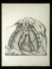
Tafel aus "Oesterreicher's anatomische Steinstiche". - Stockfleckig. - Résumé: Plate of "Oesterreicher's anatomische Steinstiche". Lithographic engraving showing the chest with heart and lung. - Foxed.
Anatomische Darstellung des Magen-Darm-Traktes.
München, (um 1828). Lithographie mit 7 Abb. Bildgrösse: 40 x 34 cm. (picture size). Blattgrösse: 54 x 40 cm. (leaf size).
Tafel aus "Oesterreicher's anatomische Steinstiche". - Stockfleckig. - Résumé: Plate of "Oesterreicher's anatomische Steinstiche". Lithographic engraving with 7 illustr. showing the gastrointestinal tract. - Foxed.
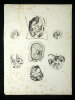
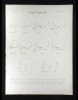
Antlitznerve. VII. Nervus facialis.
München, 1828. 2 Lithographien nach (Johann Friedrich) Meckel (dv. 1 in Umrissen). Bildgrösse: 26 x 22 cm. (picture size). Blattgrösse: 54 x 40 cm. (leaf size).
Anatomische Darstellung des "Nervus fascialis" (7. Hirnnerv). Heft XII, Tafel 1 u. 2 aus "Oesterreicher's anatomische Steinstiche. 6. Abtheilung: Nerven des menschlichen Körpers". - Stockfleckig. - Résumé: Part XII, plate 1 and 2 of "Oesterreicher's anatomische Steinstiche...". 2 lithographic engravings after Meckel showing the facial nerves wich is the seventh cranial nerve (thereof 1 in outlines). - Foxed.
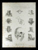
Augenhoehlennerven.
München, (um 1828). 2 Lithographien mit je 4 Abb. nach (Jules-Germain) Cloquet, (Johann Christian) Reil, (Giovanni Domenico) Santorini und (Johann Gottfried) Zinn (dv. 1 in Umrissen). Bildgrösse: 29 x 28 cm. (picture size). Blattgrösse: 54 x 40 cm. (leaf size).
Anatomische Darstellung der Augennerven. Heft X, Tafel 3 u. 4 aus "Oesterreicher's anatomische Steinstiche. 6. Abtheilung: Nerven des menschlichen Körpers". - Stockfleckig. - Résumé: Part X, plate 3 and 4 of "Oesterreicher's anatomische Steinstiche...". 2 lithographic engravings, each with 4 illustr. after Cloquet, Reil, Santorini and Zinn showing the optical nerves (thereof 1 in outlines). - Foxed.
Baender der Hand.
München, 1829. Lithographie mit 9 Abb. Bildgrösse: 42 x 30 cm. (picture size). Blattgrösse: 54 x 40 cm. (leaf size).
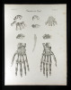
Anatomische Darstellung der Bänder der Hand. Heft VIII, Tafel 4 aus "Oesterreicher's anatomische Steinstiche. 2. Abtheilung: Bänder des menschlichen Körpers". - Stockfleckig. - Résumé: Part VIII, plate 4 of "Oesterreicher's anatomische Steinstiche...". Lithographic engraving with 9 illustr. showing the anatomy of the hand ligaments. - Foxed.
Baender des Arms.
München, 1829. Lithographie mit 9 Abb. Bildgrösse: 32 x 38 cm. (picture size). Blattgrösse: 54 x 40 cm. (leaf size).
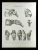
Anatomische Darstellung der Bänder des Armes. Heft VIII, Tafel 3 aus "Oesterreicher's anatomische Steinstiche. 2. Abtheilung: Bänder des menschlichen Körpers". - Stockfleckig. - Résumé: Part VIII, plate 3 of "Oesterreicher's anatomische Steinstiche...". Lithographic engraving with 9 illustr. showing the anatomy of the arm ligaments. - Foxed.
Baender des Beckens.
München, 1829. Lithographie mit 6 Abb. Bildgrösse: 43 x 30 cm. (picture size). Blattgrösse: 54 x 40 cm. (leaf size).
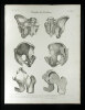
Anatomische Darstellung der Bänder des Beckens. Heft VIII, Tafel 5 aus "Oesterreicher's anatomische Steinstiche. 2. Abtheilung: Bänder des menschlichen Körpers". - Leicht stockfleckig. - Résumé: Part VIII, plate 5 of "Oesterreicher's anatomische Steinstiche...". Lithographic engraving with 6 illustr. showing the anatomy of pelvis ligaments. - Slightly foxed.
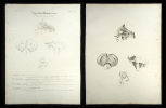
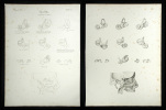
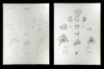
Baender des Kopfes.
München, 1829. Lithographie mit 11 Abb. nach (Jules-Germain) Cloquet, (Friedrich Heinrich) Loschge, (Joseph) Scherer und (Josias W.) Weitbrecht. Bildgrösse: 39 x 35 cm. (picture size). Blattgrösse: 54 x 40 cm. (leaf size).
Anatomische Darstellung der Bänder des Kopfes. Heft III, Tafel 6 aus "Oesterreicher's anatomische Steinstiche. 2. Abtheilung: Bänder des menschlichen Körpers". - Stärker stockfleckig. - Résumé: Part III, plate 6 of "Oesterreicher's anatomische Steinstiche...". Lithographic engraving with 11 illustr. after Cloquet, Loschge, Scherer and Weitbrecht showing the anatomy of the head ligaments. - Foxed.
Baender des Rumpfes.
München, 1829. Lithographie mit 8 Abb. Bildgrösse: 39 x 33 cm. (picture size). Blattgrösse: 54 x 40 cm. (leaf size).
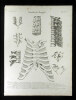
Anatomische Darstellung der Bänder des Rumpfes. Heft VIII, Tafel 2 aus "Oesterreicher's anatomische Steinstiche. 2. Abtheilung: Bänder des menschlichen Körpers". - Stockfleckig. - Résumé: Part VIII, plate 2 of "Oesterreicher's anatomische Steinstiche...". Lithographic engraving with 8 illustr. showing the anatomy of the trunk ligaments. - Foxed.
Bauchfell, Gekroes, Netze.
München, (um 1829). 2 Lithographien mit je 2 Abb. nach (Robert) Froriep (dv. 1 in Umrissen). Bildgrösse: 38 x 19 cm. (picture size). Blattgrösse: 54 x 40 cm. (leaf size).
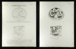
Anatomische Darstellung des Peritoneums und des Mesenteriums. Heft XX, Tafel 5 u. 6 aus "Oesterreicher's anatomische Steinstiche. 4. Abtheilung: Eingeweide des menschlichen Körpers". - Stockfleckig. - Résumé: Part XX, plate 5 and 6 of "Oesterreicher's anatomische Steinstiche...". 2 lithographic engravings with 2 illustr. after Froriep showing peritoneum and mesentery anatomy (thereof 1 in outlines). - Foxed.
Bauchhöhle.
München, 1829. Lithographie mit 7 Abb. (in Umrissen). Bildgrösse: 40 x 33,5 cm. (picture size). Blattgrösse: 54 x 40 cm. (leaf size).
Anatomische Darstellung der Bauchhöhle. Heft XX, Tafel 3 aus "Oesterreicher's anatomische Steinstiche. 4. Abtheilung: Eingeweide des menschlichen Körpers". - Stockfleckig. - Résumé: Part XX, plate 3 of "Oesterreicher's anatomische Steinstiche...". Lithographic engraving with 7 illustr. showing the abdominal cavity anatomy (in outlines). - Foxed.
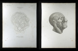
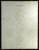
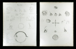
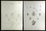
Camper's Gesichtswinkel.
München, 1829. Lithographie mit 10 Abb. nach (Peter) Camper. Bildgrösse: 30 x 34 cm. (picture size). Blattgrösse: 54 x 40 cm. (leaf size).
Vergleichende anatomische Schädeldarstellungen verschiedener Ethnien. Heft IX, Tafel 2 aus "Oesterreicher's anatomische Steinstiche. 1. Abtheilung: Knochen des menschlichen Körpers". - Leicht stockfleckig. - Résumé: Part IX, plate 2 of "Oesterreicher's anatomische Steinstiche...". Lithographic engraving with 10 illustr. after Camper showing the comparative anatomy of cranii for different ethnic groups. - Slightly foxed.
Cephalogenesis.
München, 1829. Lithographie mit 9 Abb. nach (Johann Baptist von) Spix. Bildgrösse: 38 x 32 cm. (picture size). Blattgrösse: 54 x 40 cm. (leaf size).
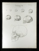
Anatomische Darstellung eines Schädels. Heft XXIV, Tafel 5 aus "Oesterreicher's anatomische Steinstiche. 1. Abtheilung: Knochen des menschlichen Körpers". - Leicht stockfleckig. - Résumé: Part XXIV, plate 6 of "Oesterreicher's anatomische Steinstiche...". Lithographic engraving with 9 illustr. after Spix showing the cranium. - Slightly foxed.
Das Auge.
München, 1829. 2 Lithographien mit 15 u. 16 Abb. nach (Samuel Thomas) Soemmerring (dv. 1 in Umrissen). Bildgrösse: 43 x 33 cm. (picture size). Blattgrösse: 54 x 40 cm. (leaf size).
Anatomische Darstellung des Auges. Heft X, Tafel 5 u. 6 aus "Oesterreicher's anatomische Steinstiche. 4. Abtheilung: Eingeweide des menschlichen Körpers". - Stockfleckig. - Résumé: Part X, plate 5 and 6 of "Oesterreicher's anatomische Steinstiche...". 2 lithographic engravings after Soemmerring, with 15 and 16 illustr. showing the eye anatomy (thereof 1 in outlines).- Foxed.
Das Ohr.
München, 1829. 2 Lithographien mit je 10 Abb. nach (Samuel Thomas) Soemmerring und (John Cunningham) Saunders (dv. 1 in Umrissen). Bildgrösse: 40 x 30 cm. (picture size). Blattgrösse: 54 x 40 cm. (leaf size).
Anatomische Darstellung des Ohrs. Heft XIV, Tafel 1 u. 2 aus "Oesterreicher's anatomische Steinstiche. 4. Abtheilung: Eingeweide des menschlichen Körpers". - Stockfleckig. - Résumé: Part XIV, plate 1 and 2 of "Oesterreicher's anatomische Steinstiche...". 2 lithographic engravings after Soemmerring and Saunders, each with 10 illustr. showing the anatomy of the ear (thereof 1 in outlines). - Foxed.
Das Ohr.
München, 1829. 2 Lithographien mit je 27 Abb. nach S(amuel) T(homas) Soemmerring (dv. 1 in Umrissen). Bildgrösse: 37 x 34 cm. (picture size). Blattgrösse: 54 x 40 cm. (leaf size).
Anatomische Darstellung des äusseren und inneren Ohres. Heft XIII, Tafel 5 u. 6 aus "Oesterreicher's anatomische Steinstiche. 4. Abtheilung: Eingeweide des menschlichen Körpers". - Stockfleckig. - Résumé: Part XIII, plate 5 and 6 of "Oesterreicher's anatomische Steinstiche...". 2 lithographic engravings after Soemmerring, each with 27 illustr. showing the ear anatomy (thereof 1 in outlines). - Foxed.
Die Nase.
München, 1829. 2 Lithographien mit 17 und 18 Abb. nach (Samuel Thomas) Soemmerring (dv. 1 in Umrissen). Bildgrösse: 42 x 30 cm. (picture size). Blattgrösse: 54 x 40 cm. (leaf size).
Anatomische Darstellung der Nase. Heft XVII, Tafel 5 u. 6 aus "Oesterreicher's anatomische Steinstiche. 4. Abtheilung: Eingeweide des menschlichen Körpers". - Leicht stockfleckig. - Résumé: Part XVII, plate 5 and 6 of "Oesterreicher's anatomische Steinstiche...". 2 lithographic engravings after Soemmerring with 17 and 18 illustr. showing the nose anatomy (thereof 1 in outlines). - Slightly foxed.